Ad monoclonal gfp antibodies for flow & icc/if. Cell surface staining of human pbmcs and cell lines.

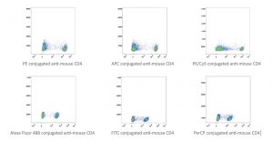
Flow Cytometric Pseudo-colorsmooth Plots Showing The Gating Strategy Download Scientific Diagram
Resuspend in facs staining buffer.

Facs buffer flow cytometry. Indirect flow cytometry (facs) protocol. • cells are measured individually but in largecells are measured individually, but in large numbers. Flow cytometry staining buffer (facs buffer) this basic facs buffer is a buffered saline solution that can be used for immunofluorescent.
Incubate on ice for 20 minutes. Run the facs instrument 🙂; Here are 5 ingredients to consider for your facs buffer:
Notes on this facs/flow cytometry methodology. Suspending cells in culture media is usually not recommended due to relatively high autofluorescence, poor ph buffering in typical flow conditions and presence of adhesion promoting components. The buffer contains sodium azide as preservative and animal serum
Another reason that people use 'protein containing buffers' for flow cytometry is to prevent cells from sticking to the side of plastic tubes (or other culture labware) as well as preventing cell clumping. Typically, the trypsin (or other detachment buffer) is quenched with culture media or a pbs/fbs buffer. Ad monoclonal gfp antibodies for flow & icc/if.
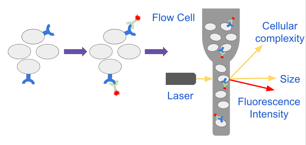
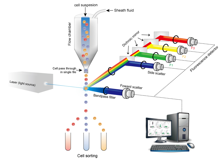
* do not add sodium azide to buffers if. Facs • flow cytometry is a technique used to measure the physical and chemical properties of cells orthe physical and chemical properties of cells or cellular components. This is problematic because it reintroduces the cations that facilitate the cells reattaching to the plate (or each other).
This is because facs is a part of the overall group of techniques called flow cytometry. Typical facs buffers consist of ca/mg free pbs with little protein (and sometimes other additives) to improve cell viability, decrease unspecific staining, and avoid sticking to plastic surfaces. The purpose of the azide in these buffers is to prevent microbial growth, but these buffers are used so quickly (and are extremely cheap to make) that you shouldn't run into any problems.
Staining protocols, antibody and cell dilution steps, wash steps required for surface staining and flow cytometric analysis. For extended storage (16 hr) as well as for. In biology, however, it is unlikely that you will use any other techniques besides this one.
*do not add sodium azide to buffers if you are concerned with recovering cell function e.g. Basic buffers for flow cytometry flow cytometry staining buffer (facs buffer) this basic facs buffer is a buffered saline solution that can be used for immunofluorescence staining protocols, antibody and cell dilution steps, wash steps required for surface staining and flow cytometric analysis. Wash 2x with biolegend's cell staining buffer and then resuspend in 0.5ml cell staining buffer for fluorescence activated cell sorting (facs), or flow cytometric analysis.
This flow cytometry staining buffer is a buffered saline solution containing fetal bovine serum and sodium azide (0.09%) as a preservative. General cell staining protocol for flow cytometry 1) except for cells grown in culture, cells obtained directly from tissues must first be resolved to a single cell suspension by means of mechanical dissociation (mincing, grinding between the ends of two frosted glass slides, dounce homogenization, steel mesh sieving, passage through Add the appropriate number of cells to be stained into a.
Basic flow cytometry staining protocol written by: Flow cytometry was performed on a bd facscan™ flowcytometry system. Flow cytometry (direct immunofluorescence staining):
Our flow cytometry staining buffer is designed for use in immunofluorescent staining protocols of cells in suspension. We recommend analysis on the same day. The system supports a wide variety of research and clinical applications and is complemented by a broad suite of intuitive software solutions to
Use of fcs or bsa in in facs buffer reduces autofluorescemce caused by non specific biding by antibodies which may falsely increase the mfi of a channel in flow cytometery cite 2 recommendations Facs may also be referred to as flow cytometry on job postings. Wash the cells twice in cold stain buffer (fbs) and pellet the cells by centrifugation (e.g., 300 x g at 4°c).
Perform red blood cell lysis, per lab protocol (either act, ack or lsm). For best results, analyze the cells on the flow cytometer as soon as possible. Flow cytometry (facs) protocols psr the bd facscalibur™ platform allows users to perform both cell analysis and cell sorting in a single benchtop system.

Pluripotent Stem Cell Flow Kit Fmc001 Rd Systems

Representative Flow-cytometric Platelet Aggregation Of Stimulated And Download Scientific Diagram
Bagaimana Memilih Antibodi Untuk Flow Cytometry - Pt Indogen Intertama

Flow Cytometry Protocols
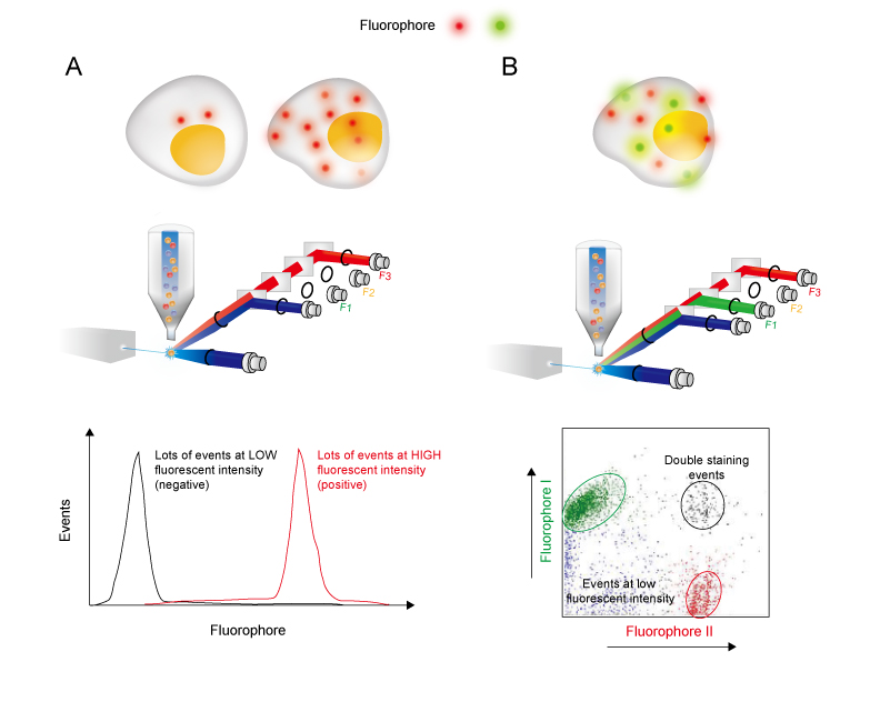
Analyzing Single Cells With Flow Cytometry
Flow Cytometry Characteristic Of Cd4 Cd25 Foxp3 Regulatory T Download Scientific Diagram

Flow Cytometry Methodology Used To Analyze Platelets Viability After Download Scientific Diagram

Flow Cytometry Facs Protocols Sino Biological
![]()
Flow Cytometry Gating Strategy For Hiv-1tg And F344 Rat Blood Download Scientific Diagram

Ldl Uptake Assay Kit Flow Cytometry Ab236208 Abcam
Flow Cytometry Guide - Creative Diagnostics

Functional Flow Cytometry Of Monocytes For Routine Diagnosis Of Innate Primary Immunodeficiencies - Journal Of Allergy And Clinical Immunology

Gating Strategy For Flow Cytometry A Strategy For Hek 293 Cells B Download Scientific Diagram

Representative Flow-cytometric Platelet Aggregation Of Stimulated And Download Scientific Diagram

Linear-and-log-scaling-in-flow-cytometry-experiments Flow Cytometry Flow Linear
Flow Cytometry Guide - Creative Diagnostics

Fundamentals Of Flow Cytometry Aat Bioquest
Flow Cytometry Guide - Creative Diagnostics

Gating Strategy Used Throughout The Experiments For Flow Cytometry Download Scientific Diagram